Файл:Red on centre green off centre.png
Перейти к навигации
Перейти к поиску
Нет версии с бо́льшим разрешением.
Red_on_centre_green_off_centre.png (634 × 474 пкс, размер файла: 48 КБ, MIME-тип: image/png)
Этот файл находится на Викискладе. Сведения о нём показаны ниже.
Викисклад — централизованное хранилище для свободных файлов, используемых в проектах Викимедиа.
|
Краткое описание
| Описание |
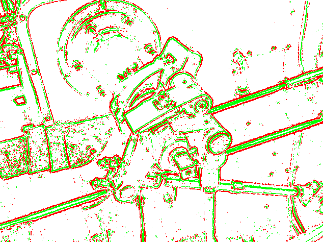
English: An example of edge detection using an emulation of retinal receptive fields. This is what the output of the optic nerve would look like (as a very rough approximation: the fovea is an almost 1:1 high definition visual area and the receptive fields increase in size from the centre of the retina to the peripheral). This image was processed in Java using a 52-pixel template (contact me for source code). Both types (on centre and off centre) cells are shown in different colours. Here is the original preprocessed image.
Русский: Распознавание границ изображения (краёв, углов) рецептивными полями сетчатки (грубая компьютерная аппроксимация). Размеры рецептивных полей увеличиваются от центра сетчатки к её периферии). Визуальная информация от двух типов клеток (с on- и off-центрами) показана красным и зелёным цветом, соответственно.
Автор изображения – User:Simpsons_contributor |
| Дата | |
| Источник | originally uploaded to the English language wikipedia |
| Автор | Собственная работа by Simpsons contributor |
Лицензирование
| Автор этого произведения, Simpsons contributor из английский Википедия, передаёт его в общественное достояние. Это разрешение действует по всему миру. В некоторых странах это не может быть возможно юридически, в таком случае: Simpsons contributor предоставляет любому право использовать данное произведение в любых целях, без каких-либо условий, если только такие условия не требуются по закону. |
Исходный журнал загрузок
The original description page was on en.wikipedia (file log). All following user names refer to en.wikipedia.
- 08:52, 31 May 2009 (UTC) Simpsons contributor 634×474 (48 KB) ({{Information |Description = An example of edge detection using an emulation of retinal receptive fields. This is what the output of the optic nerve would look like (as a very rough approximation: the fovea is an almost 1:1 high definition visual area)
История файла
Нажмите на дату/время, чтобы посмотреть файл, который был загружен в тот момент.
| Дата/время | Миниатюра | Размеры | Участник | Примечание | |
|---|---|---|---|---|---|
| текущий | 08:51, 21 марта 2011 | | 634 × 474 (48 КБ) | JeanneMish | {{Information |Description ={{en|1=An example of edge detection using an emulation of retinal receptive fields. This is what the output of the optic nerve would look like (as a very rough approximation: the fovea is an almost 1:1 high definition visual |
Использование файла
Следующая страница использует этот файл:
Глобальное использование файла
Данный файл используется в следующих вики:
- Использование в en.wikipedia.org
- Использование в fr.wikipedia.org