Файл:Brain - Lobes.png
Перейти к навигации
Перейти к поиску
Нет версии с бо́льшим разрешением.
Brain_-_Lobes.png (701 × 487 пкс, размер файла: 360 КБ, MIME-тип: image/png)
Этот файл находится на Викискладе. Сведения о нём показаны ниже.
Викисклад — централизованное хранилище для свободных файлов, используемых в проектах Викимедиа.
|
| Описание |
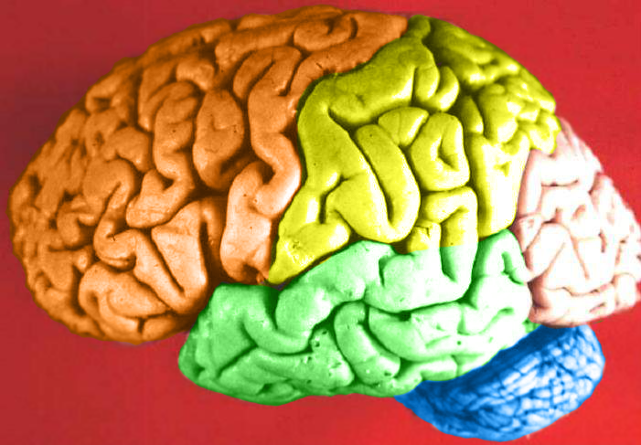
Human brain lateral view - Lobes
|
| Дата | (UTC) |
| Источник | Human_brain_lateral_view_description.JPG |
| Автор | Dep't. of Cellular Biology & Anatomy, Louisiana State University Health Sciences Center Shreveport |
| Права (Повторное использование этого файла) |
CC-BY |
| Другие версии |
| Это отретушированное изображение, что означает, что первоначальная версия изображения была изменена цифровым способом. Изменения: Hemispheres in color.. Оригинал доступен по ссылке: Human brain lateral view description.JPG:
|
Лицензирование
Я, владелец авторских прав на это произведение, добровольно публикую его на условиях следующей лицензии:
Этот файл доступен по лицензии Creative Commons Attribution 2.5 Generic
- Вы можете свободно:
- делиться произведением – копировать, распространять и передавать данное произведение
- создавать производные – переделывать данное произведение
- При соблюдении следующих условий:
- атрибуция – Вы должны указать авторство, предоставить ссылку на лицензию и указать, внёс ли автор какие-либо изменения. Это можно сделать любым разумным способом, но не создавая впечатление, что лицензиат поддерживает вас или использование вами данного произведения.
The following refers to the original source file, not this derivative version.
Этот файл, изначально опубликованный на внешнем сайте https://web.archive.org/web/20110514023714/http://www.healcentral.org/healapp/showMetadata?metadataId=40566, был проверен 1 ноября 2013 проверяющим Avenue, подтвердившим, что файл был там доступен в ту дату на условиях указанной лицензии.
|
Исходный журнал загрузок
This image is a derivative work of the following images:
- File:Human_brain_lateral_view_description.JPG licensed with Cc-by-2.5
- 2006-06-20T13:58:22Z Patho 701x487 (50176 Bytes) {{Information| |Description='''Human brain lateral view - Lobes''' # Lobus frontalis # Lobus parietalis # Lobus temporalis # Lobus occipitalis # Sulcus lateralis # Sulcus centralis # Sulcus parietooccipitalis # Incisura preo
- 2006-06-20T13:54:13Z Patho 701x487 (49891 Bytes) Auf eine alte Version zurückgesetzt
- 2006-06-20T13:51:38Z Patho 701x487 (50074 Bytes) {{Information| |Description='''Human brain lateral view - Lobes''' # Lobus frontalis # Lobus parietalis # Lobus temporalis # Lobus occipitalis # Sulcus lateralis # Sulcus centralis # Sulcus parietooccipitalis # Incisura preo
- 2006-06-20T13:28:44Z Patho 701x487 (49891 Bytes) {{Information| |Description='''Human brain lateral view''' # Lobus frontalis # Lobus parietalis # Lobus temporalis # Lobus occipitalis # sulcus lateralis # Sulcis centralis # Sulcus parietooccipitalis # Incisura preoccipital
Uploaded with derivativeFX
История файла
Нажмите на дату/время, чтобы посмотреть файл, который был загружен в тот момент.
| Дата/время | Миниатюра | Размеры | Участник | Примечание | |
|---|---|---|---|---|---|
| текущий | 22:11, 23 февраля 2009 | | 701 × 487 (360 КБ) | DavoO | {{Information |Description='''Human brain lateral view - Lobes''' # Lobus frontalis # Lobus parietalis # Lobus temporalis # Lobus occipitalis # Sulcus lateralis # Sulcus centralis # Sulcus parietooccipitalis # Incisura preoccipitalis # Polus frontalis # |
Использование файла
Следующая страница использует этот файл:
Глобальное использование файла
Данный файл используется в следующих вики:
- Использование в cs.wikipedia.org
- Использование в en.wikipedia.org
- Talk:Alcohol intoxication
- Talk:LSD
- Talk:Scopolamine
- Talk:Qigong
- Talk:Recreational drug use
- Talk:Psilocybin
- Talk:Phenomenology (philosophy)
- Talk:Alcohol (chemistry)
- Talk:Timothy Leary
- Talk:Psilocybe cubensis
- Talk:Nitrous oxide
- Talk:Atropine
- Talk:Out-of-body experience
- Talk:The Doors of Perception
- Talk:Carlos Castaneda
- Talk:Ganzfeld experiment
- Talk:Meditation
- Talk:Zen
- Talk:Hysteria
- Talk:Peyote
- Talk:Hashish
- Talk:Ketamine
- Talk:Coca
- Talk:Hippie
- Talk:Spirituality
- Talk:Mantra
- Talk:N,N-Dimethyltryptamine
- Talk:Amanita muscaria
- Talk:Hookah
- Talk:Autogenic training
- Talk:Psychonautics
- Talk:Hypnosis
- Talk:Dipropyltryptamine
- Talk:Ergot
- Talk:Phencyclidine
- Talk:Anadenanthera peregrina
- Talk:Shamanism
- Talk:Mescaline
- Talk:Diphenhydramine
- Talk:Salvinorin A
- Talk:Human Potential Movement
- Talk:Argyreia nervosa
- Talk:Terence McKenna
- Talk:Psilocybin mushroom
- Talk:Atropa belladonna
- Talk:Hypnagogia
- Talk:Kundalini yoga
- Talk:DiPT
- Talk:Dreamachine
Просмотреть глобальное использование этого файла.