Файл:Chlamydomonas TEM 09.jpg
Перейти к навигации
Перейти к поиску

Размер этого предпросмотра: 751 × 600 пкс. Другие разрешения: 301 × 240 пкс | 601 × 480 пкс | 961 × 768 пкс | 1280 × 1023 пкс | 1800 × 1438 пкс.
Исходный файл (1800 × 1438 пкс, размер файла: 784 Кб, MIME-тип: image/jpeg)
Этот файл находится на Викискладе. Сведения о нём показаны ниже.
Викисклад — централизованное хранилище для свободных файлов, используемых в проектах Викимедиа.
|
| Описание |
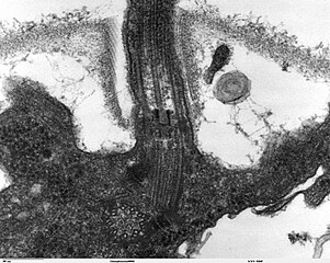
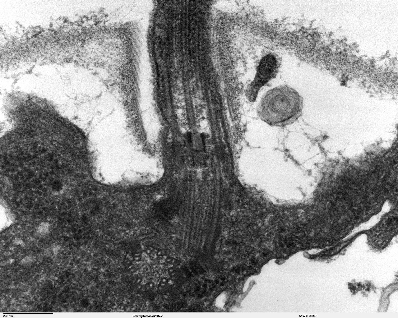
Transmission electron microscope image, showing an example of green algae (Chlorophyta). Chlamydomanas reinhardtii is a unicellular flagellate used as a model system in molecular genetics work and flagellar motility studies. This image is a longitudinal section through the flagella area. In the cell apex is the basal body that is the anchoring site for a flagella. Basal bodies originate from and have a substructure similar to that of centrioles, with nine peripheral microtubule triplets(see structure at bottom center of image). The two inner microtubules of each triplet in a basal body become the two outer doublets in the flagella. This image also shows the transition region, with its fibers of the stellate structure. The top of the image shows the flagella passing through the cell wall. |
| Дата | |
| Источник | Source and public domain notice at http://remf.dartmouth.edu/imagesindex.html |
| Автор | Dartmouth Electron Microscope Facility, Dartmouth College |
| Права (Повторное использование этого файла) |
Released into the public domain |
| Автор этого произведения, Dartmouth Electron Microscope Facility, Dartmouth College, передаёт его в общественное достояние. Это разрешение действует по всему миру. В некоторых странах это не может быть возможно юридически, в таком случае: Dartmouth Electron Microscope Facility, Dartmouth College предоставляет любому право использовать данное произведение в любых целях, без каких-либо условий, если только такие условия не требуются по закону.
|
История файла
Нажмите на дату/время, чтобы посмотреть файл, который был загружен в тот момент.
| Дата/время | Миниатюра | Размеры | Участник | Примечание | |
|---|---|---|---|---|---|
| текущий | 06:47, 21 сентября 2007 | | 1800 × 1438 (784 Кб) | Neil916 | {{Information |Description= Transmission electron microscope image, showing an example of green algae (Chlorophyta). <br><br>''Chlamydomanas reinhardtii'' is a unicellular flagellate used as a model system in molecular genetics work and flagellar motilit |
Использование файла
Следующая страница использует этот файл:
Глобальное использование файла
Данный файл используется в следующих вики:
- Использование в ar.wikipedia.org
- Использование в bs.wikipedia.org
- Использование в ca.wikipedia.org
- Использование в cs.wikipedia.org
- Использование в de.wikipedia.org
- Использование в de.wikibooks.org
- Использование в en.wikipedia.org
- Использование в es.wikipedia.org
- Использование в gl.wikipedia.org
- Использование в id.wikipedia.org
- Использование в ja.wikipedia.org
- Использование в ko.wikipedia.org
- Использование в pl.wikipedia.org
- Использование в sv.wikipedia.org
- Использование в tr.wikipedia.org
- Использование в uk.wikipedia.org
- Использование в zh.wikipedia.org