Файл:Classification and dissection of the insula.png
Перейти к навигации
Перейти к поиску

Размер этого предпросмотра: 800 × 527 пкс. Другие разрешения: 320 × 211 пкс | 640 × 422 пкс | 1024 × 675 пкс | 1280 × 843 пкс | 1935 × 1275 пкс.
Исходный файл (1935 × 1275 пкс, размер файла: 2,38 МБ, MIME-тип: image/png)
Этот файл находится на Викискладе. Сведения о нём показаны ниже.
Викисклад — централизованное хранилище для свободных файлов, используемых в проектах Викимедиа.
|
Краткое описание
| Описание |
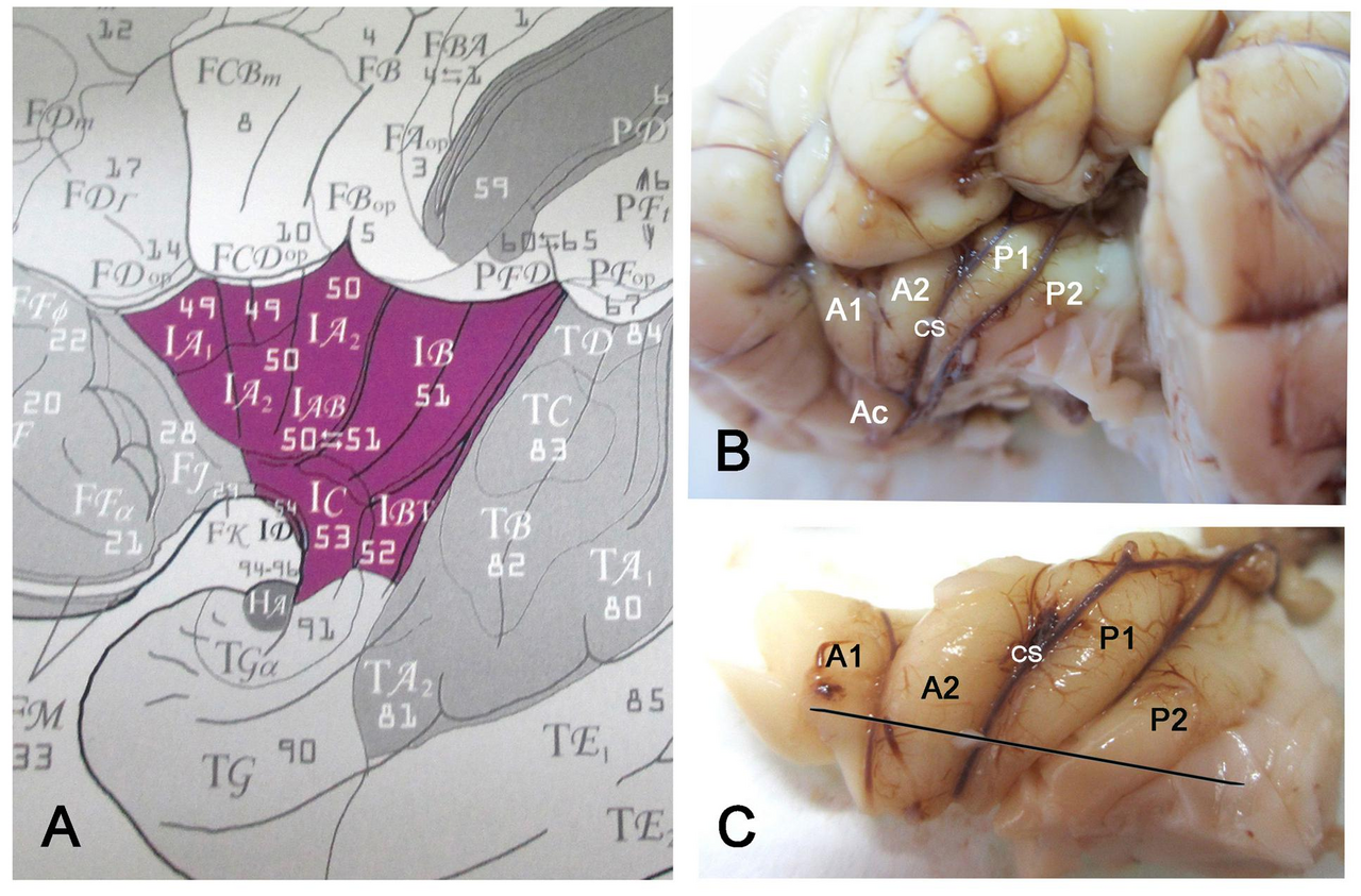
English: (A) Classification of the insular areas of Von Economo and Koskinas (1925). IA, anterior agranular, IB: posterior granular, IAB intermediate dysgranular areas. (B) Left insular lobe of a newborn infant (40 GW) after removing the anterior temporal pole. A1, A2, anterior short gyri; Ac: accessory short gyrus; P1, P2, posterior long gyri; CS: central sulcus of the insula. (C) Dissection of the insula in B. The line indicates the plane of section. |
| Дата | Published: 05 December 2017 |
| Источник | González-Arnay E, González-Gómez M and Meyer G (2017) A Radial Glia Fascicle Leads Principal Neurons from the Pallial-Subpallial Boundary into the Developing Human Insula. Front. Neuroanat. 11:111. https://doi.org/10.3389/fnana.2017.00111 |
| Автор | Emilio González-Arnay, Miriam González-Gómez and Gundela Meyer |
Лицензирование
Этот файл доступен по лицензии Creative Commons Attribution 4.0 International
- Вы можете свободно:
- делиться произведением – копировать, распространять и передавать данное произведение
- создавать производные – переделывать данное произведение
- При соблюдении следующих условий:
- атрибуция – Вы должны указать авторство, предоставить ссылку на лицензию и указать, внёс ли автор какие-либо изменения. Это можно сделать любым разумным способом, но не создавая впечатление, что лицензиат поддерживает вас или использование вами данного произведения.
История файла
Нажмите на дату/время, чтобы посмотреть файл, который был загружен в тот момент.
| Дата/время | Миниатюра | Размеры | Участник | Примечание | |
|---|---|---|---|---|---|
| текущий | 09:36, 2 июня 2019 | | 1935 × 1275 (2,38 МБ) | Was a bee | {{Information |Description={{en|1=(A) Classification of the insular areas of Von Economo and Koskinas (1925). IA, anterior agranular, IB: posterior granular, IAB intermediate dysgranular areas. (B) Left insular lobe of a newborn infant (40 GW) after removing the anterior temporal pole. A1, A2, anterior short gyri; Ac: accessory short gyrus; P1, P2, posterior long gyri; CS: central sulcus of the insula. (C) Dissection of the insula in B. The line indicates the plane of section.}} |Source=Gonzá... |
Использование файла
Нет страниц, использующих этот файл.