Файл:Maturation of the lymphatic system.jpg
Перейти к навигации
Перейти к поиску

Размер этого предпросмотра: 800 × 450 пкс. Другие разрешения: 320 × 180 пкс | 640 × 360 пкс | 1024 × 576 пкс | 1280 × 720 пкс | 2560 × 1440 пкс | 4000 × 2250 пкс.
Исходный файл (4000 × 2250 пкс, размер файла: 749 КБ, MIME-тип: image/jpeg)
Этот файл находится на Викискладе. Сведения о нём показаны ниже.
Викисклад — централизованное хранилище для свободных файлов, используемых в проектах Викимедиа.
|
Краткое описание
| Описание |
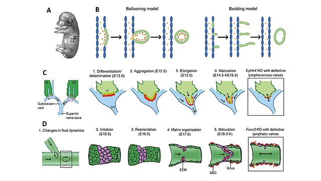
English: At late gestation and postnatally, the lymphatic vessels disconnect from the venous system and the primitive lymphatic plexus remodels into initial capillaries and collecting vessels, to finally form a functional lymphatic vasculature. These processes will be finely orchestrated by e.g. EPHB4, FOXC2, GATA2, PIEZO1, FAT4 and gap junctions. Although the lymphatics in the 5.5cm pig embryo (A) have not yet developed valves, it is apparent how the primary lymphatic plexus is gradually spreading over the entire body and invades the skin. (B) Proposed models for the disconnection of the primary lymph sacs from the venous system. The ballooning model suggests that the PROX1-expressing cells (green) in the anterior wall of the cardinal vein (blue) wall undergo a process known as delamination, like the inflation of a balloon, to form the primitive lymph sacs. As the LEC progenitors balloon out they begin to express podoplanin, critical for the binding and aggregation of circulating platelets (orange) to form a clot that will mark the disconnection between systems; completed by E14 in mice. The budding model suggests that the LEC progenitors migrate out from the venous system as strings before coalescing to form lymph sacs, maintaining venous wall integrity, and preventing vascular leakage. (C) Disconnection of the lymphatic system in the jugular region by the development of lymphovenous valves. At the sites where the lymph sacs (LS) interact with the jugulo-subclavian vein junctions, lymphovenous valve (LVV) development begins. 1. LECs (yellow) and venous endothelial cells (VECs, red) interact to form the LVV. Mechanistically, LECs upregulate the expression of Prox1, Vegfr3 and Cx43 whereas VECs upregulate the expression of Foxc2, Gata2 and Cx37. 2. The aggregate of endothelial cells begins to delaminate and reorientate itself, invaginating into the vein. 3. As the LVV develops, the cells elongate and align themselves to the direction of venous blood flow. 4. An opening in the middle of the aggregate develops and the valves recruit mural cells (orange) into the space between the valvular endothelial cells and the LECs. In mice deleted for Ephb4 (boxed-in example) lymphovenous valve formation does not go through its natural progression ending up with defective valves which cannot prevent retrograde flow. As a result, the Ephb4 knockout (KO) embryos have blood-filled lymphatics. Arrows indicate direction of fluid flow. (D) Development of secondary lymphatic valves in the collector vessels. The presence of intraluminal lymphatic valves in the collecting vessels, will ensure the unidirectional flow of lymph through the mature lymphatic system. 1. Prior to the initiation of secondary lymphatic valve development, bifurcations in the developing lymphatic network lead to changes in lymph flow re-circulation (curved arrows). 2. In these specific sites (purple area), valve formation initiates by activation of mechanotransduction pathways triggering a cascade of transcription factor upregulation (e.g. Prox1, Foxc2, Gata2). 3. The flow-induced changes in molecular identity lead the cells to change shape and reorientate. 4. Extra-cellular matrix (ECM, red) is deposited, promoting the formation of a ring-like constriction and some of the cells start protruding toward the vessel lumen. 5. During maturation, those protrusions elongate, and a sinus develops. Smooth muscle cells (SMC) cover the collectors except in the valve region. In Foxc2 knockout (KO) mice (boxed-in example), lymphatic collector vessel maturation is impaired with malformed valves. Arrows indicate bi-directional lymph flow due to reflux.
(Image credit: (A) Adapted ‘The lymphatic system in the skin of a pig 5.5cm long’ by F. Sabin, Wistar Institute of Anatomy and biology. Association of American Anatomists (1904). The American Journal of Anatomy). |
| Дата | |
| Источник | Собственная работа |
| Автор | SGUL lymres |
Sif Nielsen and eLearning Unit members Sheetal Kavia and Dhillon Khetani from St George’s, University of London (SGUL) have assisted with figure preparation. Image credit: (A) Adapted ‘The lymphatic system in the skin of a pig 5.5cm long’ by F. Sabin, Wistar Institute of Anatomy and biology. Association of American Anatomists (1904). The American Journal of Anatomy.
Лицензирование
Я, владелец авторских прав на это произведение, добровольно публикую его на условиях следующей лицензии:
Этот файл доступен по лицензии Creative Commons Attribution-Share Alike 4.0 International
- Вы можете свободно:
- делиться произведением – копировать, распространять и передавать данное произведение
- создавать производные – переделывать данное произведение
- При соблюдении следующих условий:
- атрибуция – Вы должны указать авторство, предоставить ссылку на лицензию и указать, внёс ли автор какие-либо изменения. Это можно сделать любым разумным способом, но не создавая впечатление, что лицензиат поддерживает вас или использование вами данного произведения.
- распространение на тех же условиях – Если вы изменяете, преобразуете или создаёте иное произведение на основе данного, то обязаны использовать лицензию исходного произведения или лицензию, совместимую с исходной.
История файла
Нажмите на дату/время, чтобы посмотреть файл, который был загружен в тот момент.
| Дата/время | Миниатюра | Размеры | Участник | Примечание | |
|---|---|---|---|---|---|
| текущий | 11:43, 3 февраля 2021 | | 4000 × 2250 (749 КБ) | SGUL lymres | Uploaded own work with UploadWizard |
Использование файла
Нет страниц, использующих этот файл.
Глобальное использование файла
Данный файл используется в следующих вики:
- Использование в nl.wikipedia.org