Файл:Vancomycin resistance.svg

Исходный файл (SVG-файл, номинально 2103 × 720 пкс, размер файла: 1,4 МБ)
Этот файл находится на Викискладе. Сведения о нём показаны ниже.
Викисклад — централизованное хранилище для свободных файлов, используемых в проектах Викимедиа.
|
Краткое описание
| Описание |
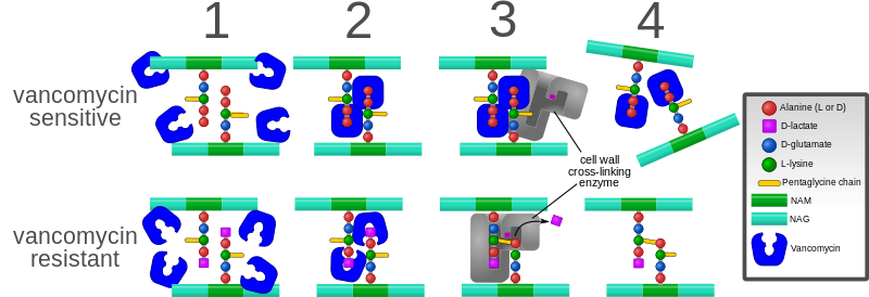
English: Diagram depicting the action of the antibiotic vancomycin and one way bacteria have evolved resistance to it.
Background: The bacterial cell wall consists of strands of repeating N-acetylglucosamine (NAG) and N-acetylmuramic acid (NAM) subunits. The NAM subunits have short peptide chains attached to them. (The exact composition of these can vary. The proximal alanine is usually L-ala and the distal two are usually D-ala.) These peptide chains are involved in forming cross-links between the strands of the cell wall. These cross-links are essential to a functioning cell wall. 1. Vancomycin is added to the bacterial environment while it is trying to synthesize new cell wall. Here, the cell wall strands have been synthesized, but not yet cross-linked. 2. Vancomycin recognizes and binds to the two D-ala residues on the end of the peptide chains. However, in resistant bacteria, the last D-ala residue has been replaced by a D-lactate, so vancomycin cannot bind. 3. In resistant bacteria, cross-links are successfully formed. However, in the non-resistant bacteria, the vancomycin bound to the peptide chains prevents them from interacting properly with the cell wall cross-linking enzyme. 4. In the resistant bacteria, stable cross links are formed. In the sensitive bacteria, cross-links cannot be formed and the cell wall falls apart. |
| Дата | |
| Источник | Собственная работа |
| Автор | Mcstrother |
| Другие версии |
[]
File:Vancomycin resistance.svg has 0 translations. |


Лицензирование
- Вы можете свободно:
- делиться произведением – копировать, распространять и передавать данное произведение
- создавать производные – переделывать данное произведение
- При соблюдении следующих условий:
- атрибуция – Вы должны указать авторство, предоставить ссылку на лицензию и указать, внёс ли автор какие-либо изменения. Это можно сделать любым разумным способом, но не создавая впечатление, что лицензиат поддерживает вас или использование вами данного произведения.
История файла
Нажмите на дату/время, чтобы посмотреть файл, который был загружен в тот момент.
| Дата/время | Миниатюра | Размеры | Участник | Примечание | |
|---|---|---|---|---|---|
| текущий | 18:27, 3 октября 2023 | 2103 × 720 (1,4 МБ) | Santanyiner | File uploaded using svgtranslate tool (https://svgtranslate.toolforge.org/). Added translation for ca. | |
| 02:08, 10 сентября 2011 | 2103 × 720 (1,65 МБ) | Mcstrother | Updated to reflect correct mechanism of cross-linking. | ||
| 14:57, 3 мая 2011 | 2103 × 720 (1,65 МБ) | Mcstrother | Changed fonts to Liberation Sans | ||
| 03:32, 10 апреля 2011 | 2103 × 720 (1,65 МБ) | Mcstrother | {{Information |Description ={{en|1=Diagram depicting the action of the antibiotic vancomycin and one way bacteria have evolved resistance to it. Background: The bacterial cell wall consists of strands of repeating N-acetylglucosamine (NAG) and N-acety |
Использование файла
Нет страниц, использующих этот файл.
Глобальное использование файла
Данный файл используется в следующих вики:
- Использование в en.wikipedia.org
- Использование в he.wikipedia.org
- Использование в sv.wikipedia.org
- Использование в zh.wikipedia.org