Файл:1gwe antipar betaSheet both.png
Перейти к навигации
Перейти к поиску

Размер этого предпросмотра: 800 × 440 пкс. Другие разрешения: 320 × 176 пкс | 640 × 352 пкс | 1024 × 563 пкс | 1280 × 704 пкс | 2000 × 1100 пкс.
Исходный файл (2000 × 1100 пкс, размер файла: 1,04 МБ, MIME-тип: image/png)
Этот файл находится на Викискладе. Сведения о нём показаны ниже.
Викисклад — централизованное хранилище для свободных файлов, используемых в проектах Викимедиа.
|
Краткое описание
| Описание |
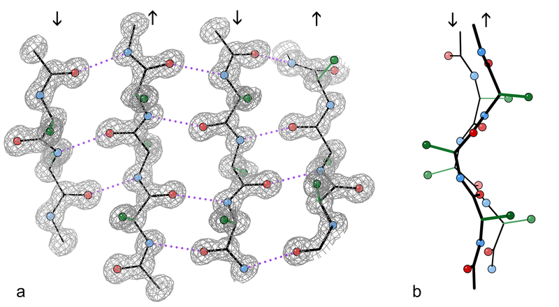
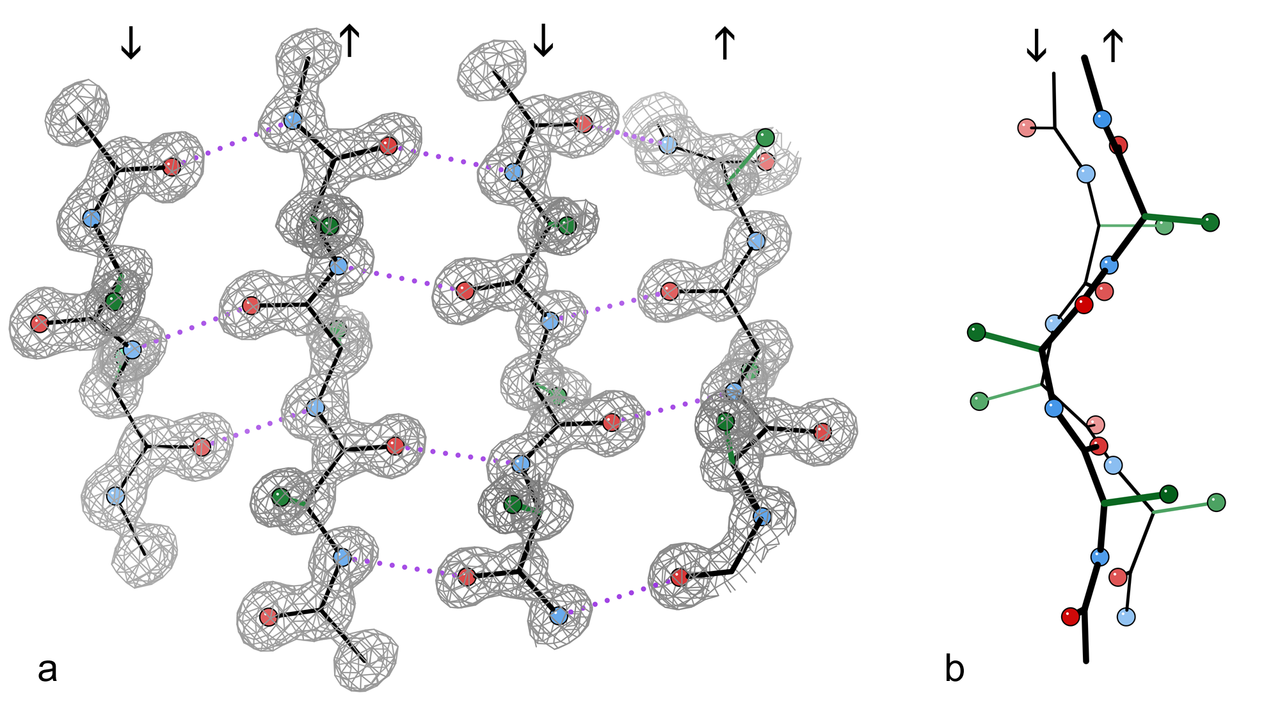
English: An example of a 4-stranded antiparallel β sheet fragment from a crystal structure of the enzyme catalase (PDB file 1GWE at 0.88 Å resolution). a) Front view, showing the antiparallel hydrogen bonds (dotted) between peptide NH and CO groups on adjacent strands. Arrows indicate chain direction, and electron density contours outline the non-H atoms. O atoms are red balls, N atoms are blue, and H atoms are omitted for simplicity; sidechains are shown only out to the first sidechain C atom (green). b) Edge-on view of the central two β strands in a, showing the righthanded twist and the pleat of Cαs and sidechains that alternately stick out in opposite directions from the sheet.
Français : Exemple d'un fragment de feuillet β à quatre chaines antiparallèles extrait de la structure cristalline de l'enzyme catalase (résolution 0,88 Å). a) Vue de face, montrant les liaisons hydrogènes (en pointillés) entre les groupes NH et CO des acides aminés adjacents. Les flèches indiquent l'orientation des chaines, et les contours de densité d'électron entourent les atomes autres que l'hydrogène. Les atomes d'oxygène sont donnés en rouge, ceux d'azote en bleu. Les atomes d'hydrogène sont omis pour plus de simplicité. Dans le même but, seul le premier carbone des radicaux est montré (en vert). b)vue par côté des deux chaines centrales montrant la torsion à droite des chaines l'une par rapport à l'autre, ainsi que les plis de chacune d'elle qui orientent les carbones portant les radicaux des acides aminés alternativement de part et d'autre de celles-ci. |
| Дата | |
| Источник | Собственная работа |
| Автор | Dcrjsr |
Лицензирование
Я, владелец авторских прав на это произведение, добровольно публикую его на условиях следующей лицензии:
Этот файл доступен по лицензии Creative Commons Attribution 3.0 Unported
- Вы можете свободно:
- делиться произведением – копировать, распространять и передавать данное произведение
- создавать производные – переделывать данное произведение
- При соблюдении следующих условий:
- атрибуция – Вы должны указать авторство, предоставить ссылку на лицензию и указать, внёс ли автор какие-либо изменения. Это можно сделать любым разумным способом, но не создавая впечатление, что лицензиат поддерживает вас или использование вами данного произведения.

|
Это изображение было оценено в соответствии с критериями ценных иллюстраций и было признано наиболее ценным изображением в категории Protein sheets and strands. Вы можете просмотреть его номинацию на странице Commons:Valued image candidates/1gwe antipar betaSheet both.png. |
История файла
Нажмите на дату/время, чтобы посмотреть файл, который был загружен в тот момент.
| Дата/время | Миниатюра | Размеры | Участник | Примечание | |
|---|---|---|---|---|---|
| текущий | 16:00, 10 апреля 2010 | | 2000 × 1100 (1,04 МБ) | Dcrjsr | {{Information |Description={{en|1=An example of a 4-stranded antiparallel β sheet fragment from a crystal structure of the enzyme catalase (PDB file 1GWE at 0.88Å resolution). a) Front view, showing the antiparallel hydrogen bonds (dotted) between pepti |
Использование файла
Следующая страница использует этот файл:
Глобальное использование файла
Данный файл используется в следующих вики:
- Использование в ar.wikipedia.org
- Использование в bg.wikipedia.org
- Использование в bs.wikipedia.org
- Использование в ca.wikipedia.org
- Использование в en.wikipedia.org
- Использование в en.wikibooks.org
- Использование в fa.wikipedia.org
- Использование в fr.wikipedia.org
- Использование в gl.wikipedia.org
- Использование в ja.wikipedia.org
- Использование в mk.wikipedia.org
- Использование в pl.wikipedia.org
- Использование в sh.wikipedia.org
- Использование в sr.wikipedia.org
- Использование в tr.wikipedia.org