Файл:Electron micrograph of neuromuscular junction (cross-section).jpg
Перейти к навигации
Перейти к поиску
Нет версии с бо́льшим разрешением.
Electron_micrograph_of_neuromuscular_junction_(cross-section).jpg (433 × 289 пкс, размер файла: 95 КБ, MIME-тип: image/jpeg)
Этот файл находится на Викискладе. Сведения о нём показаны ниже.
Викисклад — централизованное хранилище для свободных файлов, используемых в проектах Викимедиа.
|
.jpg?uselang=ru)
Краткое описание
| Описание |
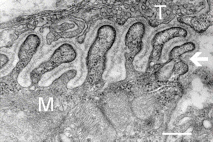
English: Electron micrograph showing a cross-section through the neuromuscular junction. T is the axon terminal, M is the muscle fiber. The arrow shows junctional folds with basal lamina. Postsynaptic densities are visible on the tips between the folds. The scale is 0.3 µm. |
| Дата | Originally uploaded to en.wikipedia on 10 марта 2006. |
| Источник | Synapse Web at the National Institute of Mental Health, National Institutes of Health; originally from en.wikipedia; description page is/was here. |
| Автор | National Institute of Mental Health; originally uploaded by Nrets at en.wikipedia. |
Лицензирование
This image is a work of the National Institutes of Health, part of the United States Department of Health and Human Services, taken or made as part of an employee's official duties. As a work of the U.S. federal government, the image is in the public domain.
 |
||
| Этот файл был определён как свободный от известных ограничений авторского права, а также связанных и смежных прав. | ||
Исходный журнал загрузок
(All user names refer to en.wikipedia)
- 2006-03-10 20:07 Nrets 433×289×8 (97758 bytes) Electron micrograph showing a cross section through the neuromuscular junction. T is the axon terminal, M is the muscle fiber. The arrow shows junctional folds with basal lamina. Postsynaptic densities are visible on the tips between the folds. Scale is 0
История файла
Нажмите на дату/время, чтобы посмотреть файл, который был загружен в тот момент.
| Дата/время | Миниатюра | Размеры | Участник | Примечание | |
|---|---|---|---|---|---|
| текущий | 03:41, 22 марта 2007 | | 433 × 289 (95 КБ) | Fran Rogers | {{Information |Description=Electron micrograph showing a cross section through the neuromuscular junction. T is the axon terminal, M is the muscle fiber. The arrow shows junctional folds with basal lamina. Postsynaptic densities are visible on the tips be |
Использование файла
Следующая страница использует этот файл:
Глобальное использование файла
Данный файл используется в следующих вики:
- Использование в ar.wikipedia.org
- Использование в cs.wikipedia.org
- Использование в de.wikipedia.org
- Использование в es.wikipedia.org
- Использование в fa.wikipedia.org
- Использование в gl.wikipedia.org
- Использование в he.wikipedia.org
- Использование в ko.wikipedia.org
- Использование в pt.wikipedia.org
- Использование в uk.wikipedia.org
- Использование в zh.wikipedia.org
.jpg)